Malignant Pleural Effusion Treatment
The pleural space is located between the lungs and the chest cavity. Normally, the expanded lung tissue causes the space to be compressed against the chest wall. Some cancers spread to this space and cause a buildup of fluid. The diagnosis of malignant pleural effusion is made by physical examination of the chest, by chest X-ray, by Cat Scan of the chest, and by analyzing the fluid that is drained from the chest. Treatment can involve drainage of the fluid from the chest, administration of medicine into the fluid, treatment of the underlying cancer, and also palliative care.
Causes
As malignant cancer fluid accumulates in the chest, the lung collapses leading to poor oxygenation and shortness of breath. Some patients may also experience chest pain, cough, and/or fevers.
Removal of the fluid in the chest cavity can provide an immediate improvement in breathing. Patients may then be able to resume their regular activities.
Treatment / Surgery
Three major options exist for the care of patients with malignant pleural effusion. They include pleurodesis, surgery, and home management with an indwelling pleural catheter.
Pleurodesis
For most patients treatment of a malignant effusion requires drainage of the fluid in the chest cavity. Typically, patients have a chest tube inserted in the involved chest cavity, which is done in the hospital. Once the fluid is drained, a “sclerosing” agent is placed into the chest cavity. The sclerosing agents cause an inflammatory reaction that obliterates the potential space in the chest cavity. When the drainage decreases, the chest tube can be removed. This treatment typically takes 7 days of hospitalization. All sclerosing agents can cause pain. The most commonly used sclerosing agent is talc, which can cause acute respiratory distress syndrome. Although not common, severe respiratory difficulties can occur leading to death.
Surgery
Videoscopic procedures have been used to manage and make the diagnosis of malignant pleural effusions. Procedures range from simply freeing collections of fluid and adding a sclerosing agent to removing the lining of the chest cavity.
Thus far there has not been any significant data demonstrating a clear advantage of surgery over a chest tube and the addition of a sclerosing agent. There is more pain than with simple chest tube insertion. Since talc is used there still is the risk of an adverse reaction to the talc. Complications such as pneumonia, infection, blood clots, and exacerbation of any pre-exsisting heart disease can also occur.
Home Management Of An Indwelling Pleural Catheter
The PLEURX® catheter by Denver BIOMEDICAL has been introduced at oncology centers nationwide as a simplified method to manage patients with malignant pleural effusions. This management strategy has been proven to relieve symptoms, improve quality of life, and decrease days of hospitalization. The following table compares the PLEURX® catheter to a standard chest tube.
| OUTCOME | PLEURX® | CHEST TUBE |
| Days of hospitalization associated with therapy | 1.5 days | 6.5 days |
| Quality of life | Equivalent | Equivalent |
| Relief of symptoms | Equivalent | Equivalent |
| Complications | Equivalent | Equivalent |
| Recurrence of effusion | 13% | 21% |
Source: Putnam JB, Light RW, et al. A randomized comparison of indwelling pleural catheter and doxycycline pleurodesis in the management of malignant pleural effusions. Cancer, 86, 1992-1999.
Other forms of surgical intervention are not necessary in these end-of-life
patients and are associated with higher morbidity, extended hospitalization,
and a 10% mortality rate.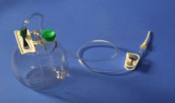
The PLEURX® catheter is a soft, supple, 16F elastic catheter with a one-way valve for safety and polyester cuff for long term placement.
The catheter is placed into the thoracic cavity under local anesthesia with minimal discomfort. This small catheter does not limit patient activity.
Drainage is performed daily using a pre-evacuated bottle and clean technique.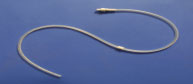
Studies have shown a very low complication rate with up to 50% of patients
achieving a spontaneous pleurodesis. When the drainage ceases, the catheter
can be removed.
Patients are admitted overnight for drainage of the effusion. The Oncology Nurse Educator instructs the patient and family member or friend very carefully on the use of the PLEURX® catheter. An instructional video supplements the education. Patients are discharged only after demonstration of proper catheter care, dressing change, drainage of fluid, and ability to document drainage on a drainage log. Medicare reimburses for the hospital stay, catheter insertion, and drainage supplies. Other insurances will need to be verified before insertion.
Many of the major cancer centers across the country have adopted the PLEURX® catheter as a first line therapy for the management of malignant pleural effusion. The University Thoracic Surgeons at RWJUH and Robert Wood Johnson Medical School also feel this should be a significant advancement for the management of this difficult disease, with a focus on palliative care.