Our Services
The GE RevolutionTM CT 256 Slice Scanner Technology in Radiology and Diagnostic Imaging
The advanced unit’s intelligent technology produces high-quality images using lower doses of radiation, contributing to more accurate diagnoses and lower exposures for patients. In addition, its speed of imaging, less than one second, is especially useful for pediatric patients who can experience imaging sedation-free, and for cardiac imaging as the whole heart can be captured in motion-free, high definition in a single beat.
The Revolution CT is designed for rapid trauma assessment, and its speed is critical when used for stroke technologies. Its use in neurology is innovative, as timing is everything when it comes to saving brain tissue. The Revolution’s applications for stroke, poly-trauma, neurology, pediatrics and cardiac CT, will ensure diagnostic confidence with the lowest level of radiation for patients.
For CT or other imaging studies or appointments, please call 973-926-7466.
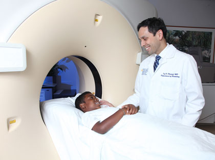
Computerized Tomography (CT) Scan
The CT Department features Phillips 16 and 64 slice CT scanners, and a GE 64 slice CT scanner equipped with low-dose ASIR (Adaptive Statistical Iterative Reconstruction). Each CT captures detailed images in seconds, allowing radiologists to reconstruct images of bone, body, brain and vascular system in 3-D. This enables physicians to diagnose conditions at their earliest stages.
We are proud to provide low-dose CT scanning with one of the few such CT scanners available in the state of New Jersey. Using advanced CT noise reduction (ASIR) we can reduce total radiation dose from a CT scan by 33-50%. This is nothing short of a miracle in dose reduction and will permit the safer imaging of obese patients and children specifically, but will also reduce the dose administered in routine cases as well.
Common CT Procedures
- CT Angiography (CTA): Non-invasive procedure used to visualize the circulatory system to diagnose vascular conditions such as peripheral vascular disease, cerebral aneurysm and pulmonary emboli. The procedure involves injecting contrast material through an IV into the blood stream.
- CT Enterography (CTE): New procedure designed to visualize the intestinal tract and surrounding organs in detail. Patients do not have to drink thick barium. The oral contrast taken prior to a CTE takes about 45 minutes to travel through the bowel and produces quality images to evaluate Crohn’s disease, inflammatory bowel syndrome and other bowel disorders.
- CT-Guided Biopsies: Under the guidance of CT imaging, abnormal tissue is sampled with precision.
- CT Body Imaging: CT imaging of the chest, abdomen and pelvis is the most common radiologic test performed to diagnose and evaluate a variety of conditions.
- CT Neuro imaging: Focuses on the diagnosis and characterization of abnormalities of nervous system, brain, spine, skull base, ENT and orbits.
Interventional Radiology: 21st Century Medicine
 NBIMC offers the region’s most comprehensive interventional radiology
services, providing patients with world-class diagnosis and treatment
from an experienced team. Our interventional radiologists use advanced
imaging techniques to diagnose conditions affecting nearly every system
of the body, and provide revolutionary, minimally invasive targeted treatments
that eliminate the need for traditional surgery for a wide range of conditions.
NBIMC offers the region’s most comprehensive interventional radiology
services, providing patients with world-class diagnosis and treatment
from an experienced team. Our interventional radiologists use advanced
imaging techniques to diagnose conditions affecting nearly every system
of the body, and provide revolutionary, minimally invasive targeted treatments
that eliminate the need for traditional surgery for a wide range of conditions.
For patients with heart disease, cancer, deep vein thrombosis, peripheral artery disease, fibroid tumors, osteoporosis and many other common medical problems, interventional radiology offers minimally invasive treatment that provide the same results as open surgery, with fewer complications and quicker recovery.
To make an appointment at our Interventional Radiology outpatient office, please call 973-926-7311.
Diagnostic Radiology
Radiography, or X-ray, is the oldest and most frequently used form of medical imaging. These images can be viewed by hospital physicians via computer, eliminating the need for film. X-ray imaging is fast and provides a rapid method of evaluating the entire body – especially the bones and chest cavity. With the exception of upper/lower GI series tests, there is no appointment necessary.
Common Diagnostic Procedures
- Chest X-ray: Performed to evaluate the lungs, heart or chest wall to diagnose pneumonia, heart failure, emphysema, lung cancer and other medical conditions.
- Upper GI Series: An upper gastrointestinal (GI) series is an X-ray or fluoroscopic examination of the esophagus, stomach and the duodenum. Patients are usually asked to swallow a solution of baking soda crystals and barium contrast liquid.
- Barium Enema: An exam to evaluate the colon and rectum during which patients are given an enema of liquid barium contrast solution. This coats the inside of the colon and rectum, producing a sharp, well-defined image. Both GI examinations are useful for diagnosing ulcers, polyps, cancer or signs of other illnesses, such as Crohn’s disease and ulcerative colitis.
- DEXA: Is the most commonly used test for measuring bone mineral density. It is the most accurate way to diagnose Osteopenia or Osteoporosis.
- Bone X-rays: Used to evaluate fractures, dislocations or other conditions.
- Hysterosalpingiograms (HSG): Images of the uterus and fallopian tubes are taken by injecting contrast via catheter inserted into the uterus. The procedure is performed by the Newark Beth Israel’s Radiologist.
Osteoporosis Detection Services (DEXA Scan)
The Breast Health Center at Newark Beth Israel Medical Center is equipped with a DEXA (Dual Energy X-ray Absorptiometry) machine, which provides bone-imaging technology. This measures bone mass and assesses an individual's risk for developing osteoporosis and its subsequent fractures. Scans are read by specially-trained nuclear physician and then the results are included in each report to the referring doctor. Physician referral is required.
Osteoporosis Detection Recommendations

Often known as a silent disease, osteoporosis can go undetected until a broken bone occurs. According to the 2004 U.S. Surgeon General's Report on Bone Health and Osteoporosis, 1.5 million older people in this country suffer fractures due to low bone density. Early detection and treatment are invaluable in helping those with osteoporosis to stay as active as possible.
Bone density testing is a safe, quick, non-invasive and painless procedure that detects osteoporosis at its earliest stages, when treatment is most effective. The current recommendation is for women to be screened by having a bone density test around the age of menopause; a repeat test every two years on the same machine is usually recommended.
To make an appointment for Osteoporosis Detection Services (DEXA Scan) call 973-926-7466.
Mammography
What Is Mammography?
Mammography is a type of imaging that uses a low-dose x-ray system to examine breasts. A mammography exam, called a mammogram, aids in the early detection and diagnosis of breast diseases in women. An x-ray (radiograph) is a noninvasive medical test that helps physicians diagnose and treat medical conditions. Imaging with x-rays involves exposing a part of the body to a small dose of ionizing radiation to produce pictures of the inside of the body. X-rays are the oldest and most frequently used form of medical imaging.
Two recent advances in mammography include digital mammography and computer-aided detection.
- Digital mammography, also called full-field digital mammography (FFDM), is a mammography system in which the x-ray film is replaced by solid-state detectors that convert x-rays into electrical signals. These detectors are similar to those found in digital cameras. The electrical signals are used to produce images of the breast that can be seen on a computer screen or printed on special film similar to conventional mammograms. From the patient's point of view, having a digital mammogram is essentially the same as having a conventional film mammogram.
- Computer-aided detection (CAD) systems use a digitized mammographic image that can be obtained from either a conventional film mammogram or a digitally acquired mammogram. The computer software then searches for abnormal areas of density, mass, or calcification that may indicate the presence of cancer. The CAD system highlights these areas on the images, alerting the radiologist to the need for further analysis.
Mammogram – Screening
This involves x-raying each breast, one from the side of the breast, and one from the top. The breast is compressed during the x-ray, improving the image by bringing the breast structure closer to the film.
Mammogram – Diagnostic
Diagnostic mammography is an x-ray exam of the breasts that is performed in order to evaluate a breast complaint or abnormality detected by physical exam or routine screening Mammogram. Diagnostic mammography is different from screening mammography in that additional views of the breast are usually taken, as opposed to two views typically taken with screening mammography.
Stereotactic Breast Biopsy
Stereotactic Breast Biopsy is a way of obtaining a sample of tissue from a questionable area that has been seen by mammography or ultrasound imaging using a special type of needle rather than surgery. Imaging by either x-ray or ultrasound will determine the exact location of area to be sampled. Your breast will be numbed prior to procedure and a needle will be placed onto the area of interest. Samples will be taken and sent to the lab for analysis. Sometimes a small clip will be placed into the breast to mark the area biopsied.
How should I prepare for a Mammogram?
Magnetic Resonance Imaging (MRI)
Our 1.5 Philips system produces clear, detailed images, to diagnose conditions at their earliest, most treatable stages. Images are interpreted by radiologists who specialize in MRI diagnosis, seven of which are fellowship-trained in neuroradiology.
Common Diagnostic Procedures
- Magnetic Resonance Angiography (MRA): A non-invasive test used to evaluate a majority of the blood vessels in the body. It can identify aneurysms, blockages and peripheral vascular disease. Images are obtained without a catheter, so there is no risk of damaging an artery.
- MRI Body Imaging: Produces high-detail images of the organs of the abdomen and pelvis.
- MRI Neuro Imaging: Produces high-detail images of the brain, spine and upper extremities.
- MRI Musculoskeletal Imaging: Produces high-detail images of major joints and soft tissues of the upper and lower extremities.
- Breast MRI: Allows for visualization of minute abnormalities that can sometimes be missed by other imaging techniques. Breast MRI is an invaluable tool in the assessment and diagnosis of breast cancer.
How should I prepare for an MRI exam?
Nuclear Medicine
The Department of Nuclear Medicine offers state-of-the-art imaging for a wide range of nuclear medicine studies. The Nuclear Medicine Department performs diagnostic scans or studies of body organs by injecting small amounts of radioactive material into a patient's veins. With the use of nuclear medicine cameras, the Department performs nearly 12,000 procedures annually.
Common Nuclear Procedures
For each of these examinations (except Thyroid Scan) a small injection is followed by a series of pictures taken by a special camera.
- Cardiac Stress Test: Evaluates adequacy of blood flow to the heart muscle.
- Bone Scan: Examines the bones for a variety of benign and malignant conditions.
- HIDA Scan: Plays a key role in determining whether gallbladder disease requiring surgery is present
- Lymphoscintigraphy: Guides the surgeon to the most important node(s) to remove in patients recently diagnosed with breast cancer or melanoma.
- Renal Scan: Assesses how well the kidneys are functioning.
- Thyroid Scan & Uptake: A pill containing a small amount of radionuclide enables the radiologist to evaluate the structure and function of the thyroid gland.
Pediatric Imaging Services
At Newark Beth Israel Medical Center and Children’s Hospital of New Jersey, pediatric imaging services include a full-range of diagnostic and interventional radiology services for newborns, infants, children, and adolescents, delivered by an expert pediatric radiology team.
Whether diagnostic imaging is needed on an emergency, inpatient or outpatient basis, our team provides gentle assurance and care that make the experience as comfortable and stress-free as possible for both children and parents.
Timely interpretation for and collaboration with our pediatricians is our priority.
Ultrasound
Common Diagnostic Procedures
- Abdominal Ultrasound: Examination of the liver, gallbladder, bile duct and spleen.
- Breast Ultrasound: Examination of the breast.
- Renal Ultrasound: Examination of the kidney.
- Gynecologic Ultrasound: Examination of the uterus and/or ovaries.
- Obstetric Ultrasound: 1st trimester examination for fetal size and due date.
- Pelvic Ultrasound: Examination of the structures and organs in the lower abdomen or pelvis.
- Ultrasound of Small Parts: Examination of the thyroid, parathyroid, parotid, scrotum and any soft tissue.
- Interventional Ultrasonography: Ultrasound-guided procedures include paracentesis; thoracentesis; liver, thyroid and renal biopsy; and fine needle aspiration of tissues. These procedures are performed on an outpatient basis. Real-time ultrasound is excellent for parotid, parathyroid, soft tissue and neck masses. Using ultrasound guidance, the radiologist is able to retrieve tissue samples for testing. Ultrasound also is used for core liver and kidney biopsies, allowing multiple tissue samples to be collected with minimal damage to surrounding tissues. Images are viewed on a screen and ultrasound guidance allows for precise needle placement.
- Ultrasound-guided Hysterosonogram: Examination of uterine and ovarian abnormalities.
- Neonatal Ultrasound: Examination of pediatric abdomen, kidney, brain, baby hips and pyloric stenosis.
Positron Emission Tomography/Computerized Tomography
PET/CT imaging combines two state-of-the-art scanner technologies into one exam. Small lesions or tumors can be detected with PET, then located with CT.
It requires a small amount of radioactive glucose to detect cancers of the breast, esophagus, cervix, lung, colon, rectum, head, neck, ovaries and thyroid, as well as melanoma and lymphoma.