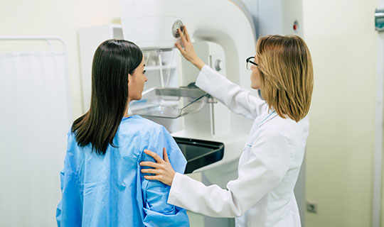
Learning you have "dense breasts" during your mammogram appointment is no reason to panic and you are not alone. Nearly half of all women who are 40 and older that get mammograms are found to have dense breast tissue on mammography.
Federal law requires that patients receive a letter indicating whether their mammogram was normal or abnormal, and New Jersey state law requires the patient be notified that their mammogram may show dense breast tissue. The state law also included a provision mandating insurance coverage for supplemental screening if breast tissue was extremely dense.
“Women with ‘extremely dense’ breasts have a higher risk of developing breast cancer than the average female population,” explained Allison Fairchild, the Clinical Breast Manager at Barnabas Health Ambulatory Care Center.
In addition, Ms. Fairchild provides the following insight regarding what it means to have dense breasts:
- Having dense breasts is perfectly normal; women’s breasts become less dense as they age.
- Dense breast tissue makes it harder for radiologists to see cancer on a mammogram.
- Having dense breasts means you have more fibroglandular tissue in your breasts than fatty tissue.
- Breast ultrasound can be considered in addition to a mammogram for those who have dense breasts.
The two main supplementary screening methods performed as complementary to a mammogram for dense breasts include whole-breast ultrasound, and contrast-enhanced breast MRI.
Dr.Sanders, Medical Director of the Breast Center, reports the added value of these two modalities compared to mammography. While mammography detects approximately 3-5 cancers per 1,000 women screened, Ultrasound can detect an additional 2-3 cancers per 1,000 women screened, and MRI has been shown to detect approximately 13-15 additional cancers per 1,000 women screened. The radiology literature is clear that MRI is the most sensitive modality for the detection of breast cancer.
While breast ultrasound is a good screening tool for women with dense breasts, it can also be used to find the cause of the breast symptoms, check a breast lump found on breast self-examination or physical examination, and further analyze certain abnormal findings seen on mammography.
Ms. Fairchild also shares what to expect during a breast ultrasound:
- The exam usually takes 30 minutes or less.
- The patient is scanned while lying on supine, face-up.
- The technologist uses a wand-like device called a transducer to make the images.
- A warm gel is used on the breast for comfort.
As mentioned, it is important to note that both US and MRI are supplementary to mammography and must be performed in conjunction with a recent mammogram. As Dr. Sanders emphasizes, neither US nor MRI can detect tiny abnormal microcalcifications that can be seen only on mammography which may indicate the earliest type of breast cancer, DCIS (ductal carcinoma in situ), which is completely curable (Stage 0).
The Breast Center at Cooperman Barnabas Medical Center, located at the Barnabas Health Ambulatory Care Center, has been committed to providing patients the imaging resources for early breast cancer detection for decades. This program has been recognized as a Breast Imaging Center of Excellence by the American College of Radiology and is accredited by the National Accreditation Program for Breast Centers.
To schedule a mammogram appointment, book online or call 855-654-5393.
For a referral to a RWJBarnabas Health primary care physician or breast specialist, call 888-724-7123.