A far less invasive surgery is now offered close to home.
There are many different types of skull base tumors, which can be either malignant or benign. What they all have in common is that they’re extremely challenging to remove because they’re so close to the brain, eyes, critical nerves and arteries.
The traditional way of removing these hard-to-reach tumors involves a craniotomy (ear-to-ear opening of the skull). Today, there’s an advanced and safer option for most cases: endoscopic endonasal skull base surgery, which employs a narrow, lighted telescope called an endoscope to reach these tumors through the nasal passage. The surgery is now available at Saint Barnabas Medical Center (SBMC).
A DELICATE OPERATION
The skull base is the floor of the cranium, the area where the brain rests in the skull. It’s made of five bones fused together. This small, intricate area includes major arteries and important nerves that pass through openings in the skull base to connect with the rest of the body.
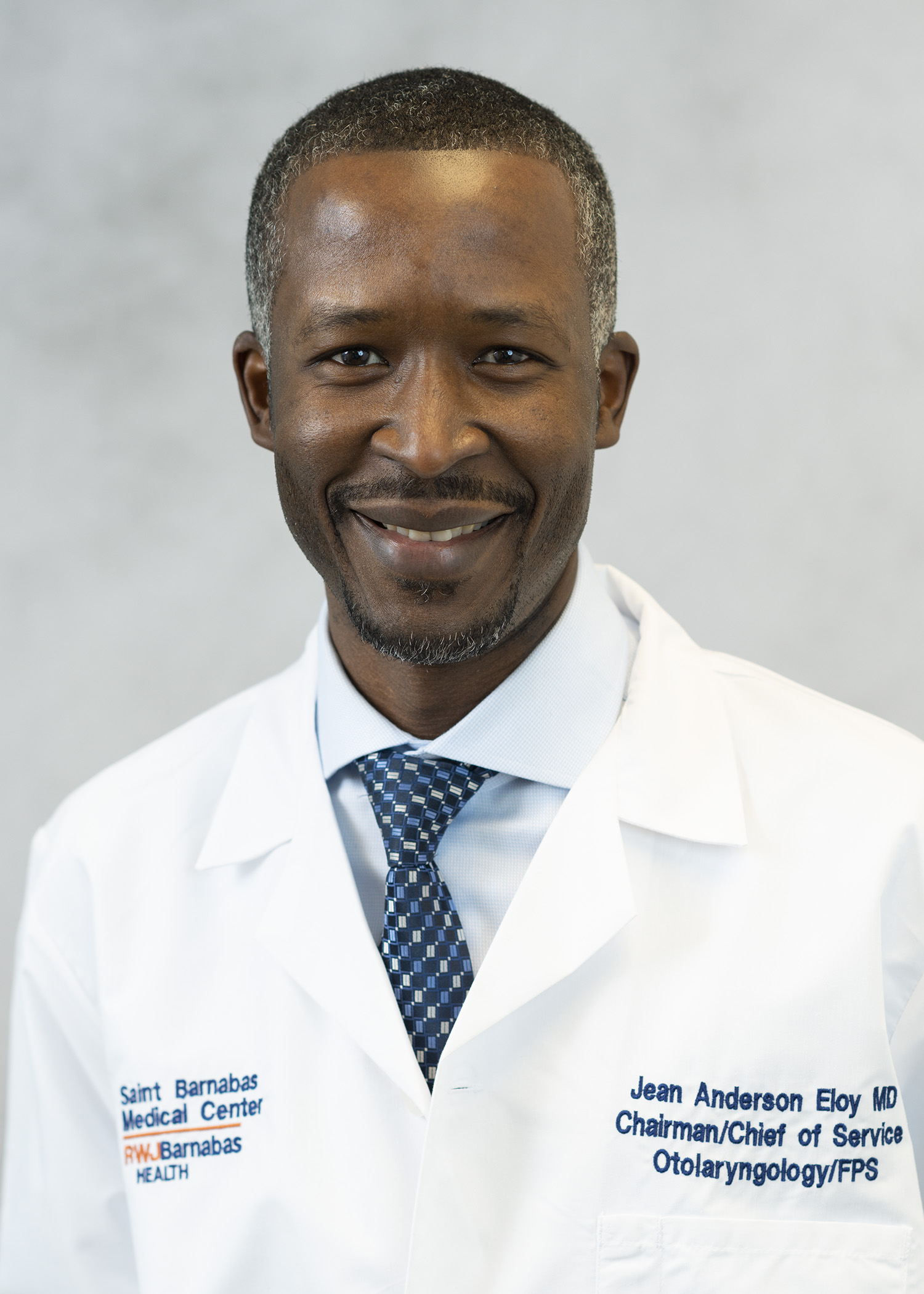
 “Tumor growths in this area include pituitary adenomas, craniopharyngiomas, meningiomas, chordomas, chondrosarcomas, as well as many types of nasal cavity and sinus benign and cancerous tumors. Other pathologies in this area include cerebrospinal fluid leaks and congenital conditions that patients are born with,” explains Jean Anderson Eloy, MD, FACS, FARS, Chair of Otolaryngology–Head and Neck Surgery at SBMC.
Endoscopic skull base surgery requires a multidisciplinary team of trained surgeons working together. “Patients need experienced doctors who do a high volume of these procedures,” says Dr. Eloy. Besides Dr. Eloy, the SBMC team includes skull base neurosurgeon James K. Liu, MD, FACS, FAANS; sinus surgeon Wayne Hsueh, MD; and ophthalmic plastic and reconstructive surgeons Paul D. Langer, MD, FACS and Roger E. Turbin, MD, FACS.
Magnetic resonance imaging (MRI) and computed tomography (CT) scans used with image guidance (a type of GPS navigation used during surgery) serve as critical guides, helping surgeons pinpoint a tumor’s location. Surgeons work with extreme precision so as not to disrupt the senses or damage critical nerves and blood vessels nearby. After the growth is removed, synthetic materials and natural tissues are used to reconstruct the skull base. (Patients with cancerous tumors may be referred to the Department of Radiation Oncology and Medical Oncology for further treatment.)
Endoscopic skull base surgery offers a quicker recovery than the traditional surgical method. Hospital stays are brief, the risk of infection is reduced and patients are typically back on their feet in days.
EXPERTISE AT HAND
Dr. Eloy founded New Jersey’s first fellowship program in Rhinology and Endoscopic Skull Base Surgery at Rutgers New Jersey Medical School. Drs. Eloy and Liu, Co-Directors of the Endoscopic Skull Base Surgery Program, have collaborated for a decade, and have among the most extensive experience in endoscopic skull base surgery in the world.
SBMC treats about 200 patients with skull base tumors each year. “Still,” says Dr. Eloy, “many New Jersey physicians and patients don’t realize we offer this specialized surgery in their backyard.”