PET/CT and Nuclear Medicine

Nuclear Medicine imaging offers early detection and is used in the diagnosis, management and treatment of serious disease, which may result in a more successful prognosis. A PET CT scan is a diagnostic tool used to reveal the cellular level metabolic changes occurring in an organ or tissue and commonly used to detect cancer, heart problems (such as sarcoidosis), brain disorders (including brain tumors, memory disorders, seizures) and other central nervous system disorders.
A PET CT scan involves injecting a very small dose of a radioactive chemical, called a radiotracer, into the arm which travels through the body and absorbed by the organs and tissues being studied. Physicians are provided with cross-sectional images of the body organ from any angle which enables them to detect any abnormal uptake.
We also offer PET CT of the Breast for ER+ lesions using agent Cerianna.
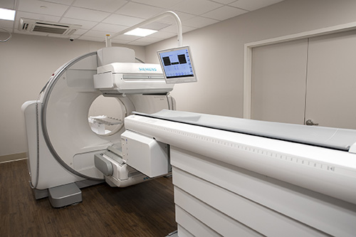
Our SPECT/CT scanner combines functional information with precise functional anatomical location. High-quality Nuclear SPECT/CT imaging can be particularly helpful for cases involving parathyroid, infectious disease and orthopedics. Recently installed, our new SPECT/CT camera provides the most up-to-date technology in the field of molecular imaging.
