The Center for Women's Health offers the latest in mammograms.
 October is the month of the pink ribbon in honor of breast cancer awareness. That ribbon is a good reminder to get a mammogram, a screening tool for breast cancer. Aside from certain types of skin cancer, breast cancer is the most common cancer in women. There’s a 1 in 8 chance that a woman will develop breast cancer at some point in her life.
Fortunately, radiologists are now able to detect very small tumors thanks to an advanced imaging technique.
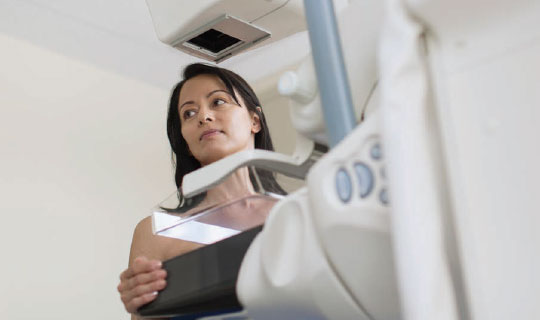
Three years ago, the Robert Wood Johnson University Hospital (RWJUH) Hamilton Center for Women’s Health began offering three-dimensional (3-D) mammography, also known as breast tomosynthesis. With this technique, breast images are sliced thinly, and 100 to 300 images are generated instead of the standard four.
“This improves our ability to detect breast cancer,” says Sandip Nayee, DO, the Center’s lead radiologist. “We look at each 'slice’ of the breast and can detect tumors that are sometimes not apparent on two-dimensional mammograms.” Breast tomosynthesis also decreases the chances of being called back for a follow-up mammogram.
Three-dimensional mammograms are able to detect small tumors more easily in dense breasts. They can also be beneficial in women with fatty breasts. Subtle areas of distortion, which may indicate an underlying tumor, are clearer on 3-D images.
Follow up testing
The Center for Women’s Health also offers diagnostic mammography, in which a followup mammogram is performed if a finding on an initial screening mammography requires additional investigation. “We want to be sure that what we saw on the initial mammogram is a persistent finding,” says Dr. Nayee. If a nodule, or lump, is found, an ultrasound—a test that uses sound waves to create images of the body—may be performed. If the nodule is a fluid-filled cyst, no further testing is needed. But a solid nodule may need to be biopsied. “Approximately 75 percent of breast biopsies are not cancer,” says Dr. Nayee.
Women at average risk for breast cancer should begin having annual mammograms at age 40. Those
who have risk factors, such as a genetic mutation or a first-degree relative with breast cancer (such as a sibling, mother or child), should be screened earlier. Talk with your physician about the right screening program for you.
To schedule a mammogram, visit www.rwjbh.org/rwjhamiltonbreast.
Help for high-risk patients
If you’re concerned about your risk for breast cancer, you may benefit from the Robert Wood Johnson University Hospital Hamilton and Rutgers Cancer Institute of New Jersey Hereditary Oncology Prevention and Evaluation (HOPE) Program. The program provides genetic counseling and testing, as well as social support.
A person has an initial consultation with a genetics counselor. If he or she is found to be at high risk, he or she can undergo formal testing, says Patty Hutman, a breast health navigator and coordinator for the HOPE Program. The person provides a saliva sample, and a chromosome analysis is performed. Results are available in seven to 14 days and are reviewed during a followup consultation. To learn more about genetic counseling, call 609.584.2836
RWJBarnabas Health and RWJUH Hamilton, in partnership with Rutgers Cancer Institute of New Jersey—the state’s only NCI-Designated Comprehensive Cancer Center—provide close-to-home access to the latest treatment and clinical trials. For more information, call 844.CANCERNJ or visit www.rwjbh.org/rwjhamiltoncancer